
CT (Computed Tomography) scan machines and MRI (Magnetic Resonance Imaging) machines are both medical imaging devices, but they use different principles and technologies to produce images of the body. Let’s discuss the difference between the MRI and CT scan machine in brief.
CT Scan (Computed Tomography)
A CT (Computed Tomography) scan machine, also known as a CT scanner or CAT (Computerized Axial Tomography) scanner, is a medical imaging device used to create detailed cross-sectional images of the body’s internal structures. It combines X-ray technology with advanced computer processing to generate these images. Here’s an overview of the components and functionality of a CT scan machine:

1. Gantry
The gantry is the large, circular structure that houses the X-ray tube and the detector array. It rotates around the patient during the scanning process to acquire images from different angles.
2. X-Ray Tube
The X-ray tube emits a narrow beam of X-rays that passes through the patient’s body. The machine’s operator controls the intensity of the X-ray beam.
3. Detector Array
The gantry locates the X-ray tube opposite the detector array. It consists of multiple detectors that measure the X-rays that pass through the patient’s body. These detectors convert the X-rays into electrical signals.
4. Patient Table
The patient table is a movable platform that the patient lies on during the scan. The gantry slides in and out, allowing the patient to position themselves correctly for the scan.
5. Computer System
The computer system processes the data from the detectors and reconstructs it into detailed cross-sectional images. The process involves complex mathematical algorithms that create detailed images of the body’s internal structures.
6. Operator Console and Control Panel
The operator console is where the technologist or radiologist controls the CT scan machine. They can adjust scan parameters, monitor the scan progress, and review the acquired image. The control panel allows the operator to set parameters such as scan duration, X-ray intensity, and the thickness of the cross-sectional slices.
8. Couch & Positioning Devices
The couch and positioning devices help ensure that the patient is correctly positioned and immobilized during the scan. This is crucial for obtaining accurate images.
9. Collimators and Display Monitors
Collimators are devices that help shape and limit the X-ray beam, focusing it on the area of interest and reducing unnecessary radiation exposure to surrounding tissues. The operator uses display monitors to monitor the scan progress in real-time and review the reconstructed images.
Some CT scans require the use of contrast agents to enhance the visibility of blood vessels, certain tissues, or abnormalities. The CT scan machine may integrate an injection system for administering these contrast agents.
Modern CT scan machines come in various designs, including single-slice, multi-slice (which can acquire multiple slices in a single rotation), and cone-beam CT for specialized applications like dental imaging. The technology has evolved to provide faster scans with lower radiation doses, making it a valuable tool for diagnosing a wide range of medical conditions.
MRI Scan (Magnetic Resonance Imaging)
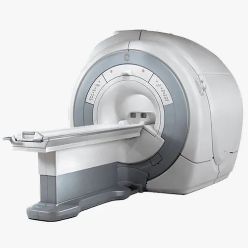
An MRI (Magnetic Resonance Imaging) machine is a sophisticated medical imaging device that uses strong magnetic fields and radio waves to create detailed images of the internal structures of the body. Here’s an overview of the components and functionality of an MRI machine:
1. Magnet System
The core of the MRI machine is its powerful magnet. This magnet generates a strong magnetic field that aligns the hydrogen nuclei (protons) within the body’s tissues. Someone measures the strength of the magnet in units called teslas (T), with higher tesla values indicating stronger magnets.
2. Gradient Coils
Gradient coils are additional coils of wire placed within the main magnet. These coils create varying magnetic fields across different spatial dimensions (x, y, and z) in order to encode spatial information. They help differentiate the location of signals within the body, which is crucial for creating detailed images.
3. Radiofrequency (RF) Coils
Radiofrequency signals are transmitted and received using RF coils. The placing of these coils around the area of the body is being imaged. The coils send RF pulses which disrupt the alignment of the hydrogen nuclei, and the returning nuclei use their emitted signals to create images as they return to their original alignment.
4. Patient Table
The patient table moves with the patient into the cylindrical bore of the MRI machine. Reposition the table during the scan to capture images of different body areas.
5. Gradient Amplifier
Gradient amplifiers provide the electrical currents needed to power the gradient coils. They allow for precise control of the gradients and enable the creation of detailed spatial images.
6. Radiofrequency (RF) System
The RF coils transmit the radiofrequency pulses generated and controlled by the RF system. These pulses manipulate the alignment of the hydrogen nuclei, allowing for the acquisition of image data.
7. Computer System & Software
The computer system processes the data acquired from the RF coils and gradient coils. Use of complex mathematical algorithms to transform the raw data into detailed images. Specialized software controls the scan parameters and sequences.
8. Operator Console
The operator console is where the MRI technologist or radiologist controls the MRI machine. They select the appropriate imaging sequence, adjust parameters, and monitor the scan progress.
9. Display Monitors
The operator uses display monitors to monitor the patient and the scan progress in real time. The reconstructed images are also displayed on these monitors.
10. Bore
The bore is the tunnel-like central opening of the MRI machine where the patient lies during the scan. The bore’s diameter can vary, with larger bore sizes designed to accommodate patients who might feel claustrophobic in narrower bores.
11. Shielding
Specialized shielding prevents external electromagnetic interference from affecting the images of MRI machines housed within rooms. The strong magnetic fields can also pose risks to individuals with certain medical implants or devices.
Modern MRI machines come in various types, including open MRI (with a more spacious design) and high-field MRI (with stronger magnets for higher image quality). MRI is a versatile imaging modality used to visualize soft tissues, organs, blood vessels, and even certain neurological functions without using ionizing radiation.
Here are the key differences between CT scans and MRI scans.
Principle of Imaging
- CT Scan: CT uses X-rays to create cross-sectional images of the body. It rotates an X-ray source around the patient, and detectors measure the X-rays that pass through the body. The data is then processed by a computer to generate detailed cross-sectional images.
- MRI: MRI uses strong magnetic fields and radio waves to create detailed images of the body’s internal structures. It does not involve ionizing radiation, making it a safer option for certain patients, such as pregnant women and young children.
Image Contrast
- CT Scan: CT is excellent for imaging dense tissues like bones and is particularly useful for detecting fractures, tumors, and other abnormalities in the chest, abdomen, and pelvis.
- MRI: MRI excels at imaging soft tissues like organs, muscles, and nerves. It is especially valuable for brain and spinal cord imaging, joint problems, and detecting abnormalities in organs like the liver and kidneys.
Image Detail
- CT Scan: CT scans provide detailed images with excellent spatial resolution, allowing for precise anatomical visualization.
- MRI: MRI produces images with superior contrast resolution, making it highly effective in differentiating between different soft tissues and detecting abnormalities that may be challenging to see with other imaging modalities.
Safety and Speed
- CT Scan: CT involves exposure to ionizing radiation, which carries a small but potential risk of adverse effects, especially with frequent scans or in younger patients. They are relatively quick, usually taking only a few minutes to complete the imaging process.
- MRI: MRI does not use ionizing radiation, making it a safer imaging option for certain individuals, although it is not recommended for patients with certain metal implants or pacemakers. MRI scans generally take longer than CT scans, often ranging from 15 minutes to over an hour, depending on the specific examination and imaging sequences required.
Radiation Exposure
- MRI: MRI does not use ionizing radiation, making it safer in terms of radiation exposure. It is generally considered safe for most patients, including pregnant women and children.
- CT Scan: CT scan involves exposure to X-rays, which can potentially pose a risk if used excessively. However, modern CT scanners use techniques to minimize radiation doses.
Patient Comfort
- MRI: Some patients may feel uncomfortable in the confined space of the MRI machine, and individuals with claustrophobia might find it challenging.
- CT Scan: The CT scanner is less confining, and many patients find it more comfortable.
Limitations
- CT Scan: CT is not as effective in distinguishing between certain soft tissues, and the use of ionizing radiation should be minimized, especially in pediatric and pregnant patients.
- MRI: MRI may be less suitable for patients who have claustrophobia, metal implants that are not MRI-safe, or other conditions that limit their ability to lie still for an extended period.
Applications
- MRI: MRI is often preferred for imaging the brain, spinal cord, joints, muscles, soft tissues, and certain tumors.
- CT Scan: CT scan is frequently used for imaging the chest, abdomen, pelvis, bones, blood vessels, and emergency situations like trauma.
Conclusion
Both MRI and CT scans have their own strengths and are chosen based on the clinical situation, the specific area of interest, the desired level of detail, and the patient’s condition. Each modality complements the other and contributes to a comprehensive diagnostic approach in modern medicine.
The choice between CT and MRI depends on the specific clinical question, the area of the body to be imaged, and the patient’s individual circumstances. In many cases, both imaging modalities complement each other, and healthcare providers may use them together to get a comprehensive view of a patient’s condition.