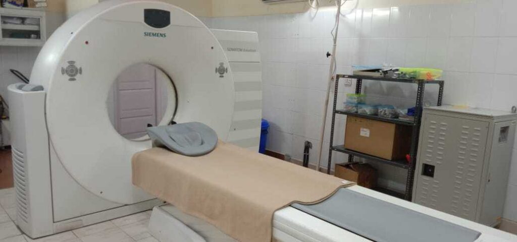
A CT (Computed Tomography) scan is an imaging procedure that uses special X-ray equipment to create detailed pictures inside the body. The images created by a CT scan show more detail and can often diagnose medical conditions earlier and more accurately.
Doctors use this information to examine organs, bones, soft tissue, and blood vessels. To diagnose illnesses or injuries much quicker and improve treatment options available for patients.
During the Scan
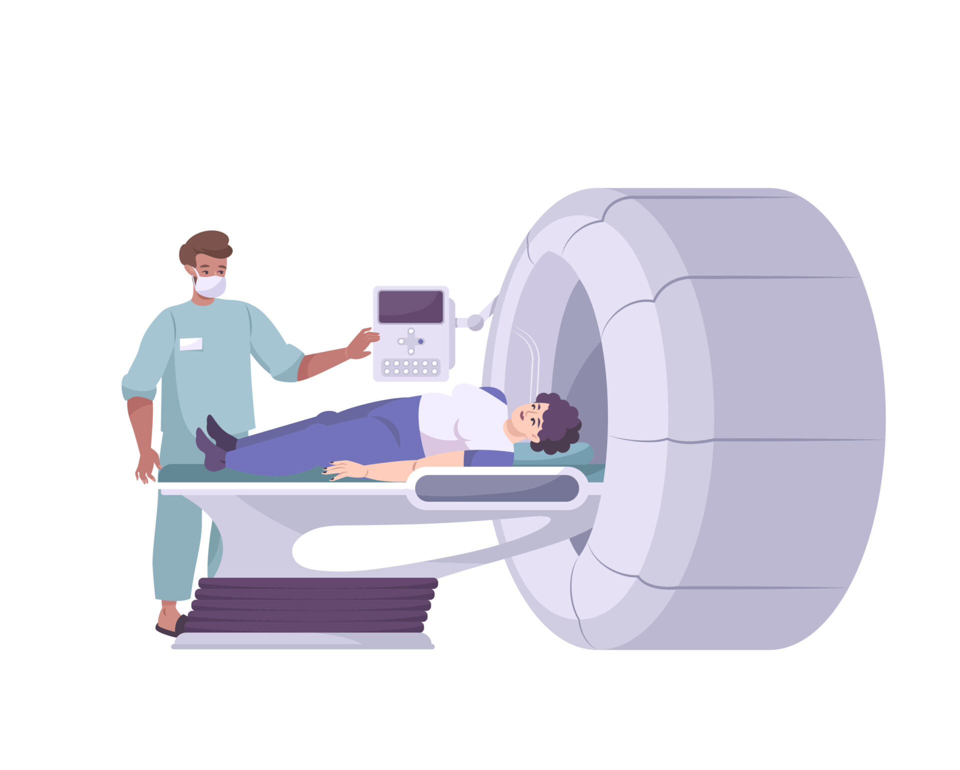
- Patients typically lie on a table that slides into a large doughnut-shaped machine.
- They will ask them to remain still while they take scans from different angles of internal organs and structures once they have settled.
- Stay as relaxed as possible – any movement or breathing too quickly can make scans blurry or distort results.
- The entire process usually lasts between 15 minutes and an hour depending upon what’s being examined.
- In addition, most clinics use a contrast dye to highlight areas that need further focus which makes patients feel discomfort.
- Radiation: (Also called a useful beam) is the radiation emitted directly from the X-ray tube that is used for patient imaging.
- Primary Barrier: It is a wall, ceiling, floor, or other structure that will intercept radiation emitted directly from the X-ray tube.
- Secondary Radiation: Consists of X-rays scattered from the patient and other objects such as imaging hardware and leakage radiation from the protective housing of the X-ray tube.
- Barrier (Secondary): It is a wall, ceiling, floor, or other structure that will intercept and attenuate leakage and scattered radiation.

Radiation Exposure
CT scans are an important diagnostic tool, used to diagnose a wide range of medical conditions. However, the benefits from such imaging can be great, there is also the potential risk associated with radiation exposure.
These exposures can add up with repeated scans or multiple different types of images acquired at once. During a standard scan, there is up to 100 times more exposure than X-rays. Also, high doses can significantly increase their risk of developing certain types of cancers later in life.
When calculating the total dose for children or pregnant women, doctors must take into account not only this radiation exposure from one single exam but also any other prior irradiations encountered in their life as well.
We are regularly exposed to various kinds of radiation like radioactive materials, radon a radioactive gas. It comes from the natural breakdown of the uranium in soil, water, rocks, building blocks, and cosmic rays from outer space.
Countries vary in their background radiation. The largest source of background radiation is radon which is typically about 2 mSv per year, a natural gas found in our homes.
Location is also important, being at a higher altitude increases a person’s exposure. Radiation we get from the CT scan machines, and X-rays are ionizing radiation.
These forms of radiation can travel through living tissue and interact with molecules to form ions. After that causes damage in cells within that area, leading to potential health risks.
Ionizing radiation can damage the cells and the tissues, and sometimes they just do the job in the wrong way. They leave the small areas neglected which results in DNA mutation which later on in future develops the cancer cell.
The average radiation one should take is 3 millisieverts per year(mSv).
The extent of potential damage depends on several factors:
- type of radiation
- sensitivity of the affected tissues and organ
- manner and length of time exposed
- radioactive isotopes involved
- characteristics of the exposed person (age, gender, weight, etc)
Methods such as radiographs or X-rays involve very low doses while other techniques like CT scans or PET scans may require higher levels of exposure.
The amount of the measure of radiation exposure depends on the imaging test and the part of the body, the absorption rate will also vary depending on an individual’s age and weight.
- A single chest X-Ray exposes the patient to about 0.1mSv. People naturally expose themselves to the same amount of radiation over about 10 days.
- Mammogram exposes a woman to about 0.4 mSv; which is equal to about 7 weeks of natural exposure.
- Lower GI (Gastrointestinal) series using x-rays of the large intestine exposes a person to about 8 mSv, or equal to the amount expected over about 3 years.
- CT scan of the abdomen (belly) and pelvis exposes a person to about 10 mSv.
- PET/CT exposes a person to about 25 mSv of radiation. This is equal to about 8 years of average background radiation exposure.
Radiation exposure can also cause acute effects such as nausea and vomiting, skin redness, hair loss, radiation burns, acute radiation syndrome, or even death.
Excessive Radiation Exposure can increase the risk of cancer, during nuclear emergencies, it releases radioactive iodine, which if swallowed or breathed can develop thyroid cancer.
Ensure Radiation Safety for patients
- Limiting the total “beam–on” time
- Avoiding oblique lateral projections
- Collimation to limited beam size
- Selecting low dose rate protocol
- Use of exposure protocols for patient examinations including pediatric patients
- The use of DLP(Dose Lenght Product)in CT and DAP(Dose Area Product) values for IR (Interventional Radiology)procedures
- record-keeping patient’s doses for CT and IR procedures.

Does extra radiation increase a person’s cancer risk?
Radiation Exposure depends on various factors like age, health, height, weight, gender, and other factors. While research has yet to provide concrete answers, many studies have linked exposure to high levels of radiation with an increased possibility of health problems.
Those who voluntarily receive high-dose radiotherapy treatments can be at higher risks for short or long-term health effects related to cancer development. While it may not always lead directly to cancer formation.
Healthcare professionals should monitor exposure to large amounts of extra radiation with frequent checkups.
There is a growing concern that extra radiation such as MRI and CT scans might increase the risk of cancer in kids. Studies have found an increased risk of central nervous system tumors. It is especially for children who have several medical imaging tests within close succession.
However, doctors can minimize this potential risk by discussing other diagnostic modalities which do not involve radiation. In addition to monitoring usage for symptoms alone, it’s important to consider any additional risks associated with repeated exposure.
It’s important to consider this information when using any form of medical imaging. Healthcare professionals should supervise overexposure carefully to avoid potentially serious consequences later on in life.
Proper safety protocols must be followed to minimize the radiation impact of CT scan machines. Exposure to radiation increases a person’s risk of developing cancer or other illnesses and has an exponential effect.
It is important for healthcare workers to understand the implications of ionizing radiation so that they can take steps to mitigate its dangers and operate these machines safely. By doing this, we can ensure that all patients get access to timely treatments without compromising their health in any way.
Understanding the impact of Radiation Exposure from a new generation CT Scan Machine.
Increased adoption of new-generation computed tomography (CT) scans for medical purposes is happening nowadays. The use of advanced technologies relies on radiation exposure to achieve images with high resolution and accuracy. Exposure Advances in technology have made it possible for modern medical imaging techniques to use less radiation than ever before.
One such technique is the new-generation computed tomography (CT) scan machine, which promises a lower risk of negative health effects from exposure to high doses of radiation.
This article explores the potential impacts that radiation has on patients or other personnel who are present during a scan procedure. As well as how much risk this poses compared to other diagnostic imaging technology.
A computed tomography (CT) scan uses X-rays from multiple angles around an object or person’s body to create detailed images inside the body. Without having organs or parts cut open surgically.
This powerful tool allows healthcare professionals to diagnose complex diseases with greater accuracy and speed. It comes with some risks due to its usage of ionizing radiation which has been linked to increased long-term cancer.
The Risk Involved: How much radiation are we exposing ourselves to?
Although modern equipment has improved significantly in terms of radiation emission reduction. There will always be some risk involved when undergoing any radiographic procedure due to its ionizing nature.
It’s important for both patients and operators to understand what doses they can expect from current innovation machines. Avoid long-term effects caused by irradiation accordingly. Therefore professional assessments need to take place before scanning someone’s body with them.
Children younger than 18 years old and pregnant women should apply precautionary methods. Also, people having chronic illnesses like cancer could suffer great harm if exposed too often to scanners.

Advantages of New Scanners
- New technology techniques such as adaptive dose control (ADC) offer greater safety benefits. When using new generations of CT scan machines they adjust the amount of X-ray energy released.
- Only give necessary doses to protect patients from excessive amounts.
- Another development is 3D scanning capabilities which greatly improve accuracy when diagnosing certain conditions without compromising patient safety.
- Furthermore, newer scanner systems may provide more comfortable positions during procedures reducing further stress. It helps scans take less time giving even lower dosage rates over prolonged periods.
- Newer generation devices offer lower amplification rates meaning radiologists can obtain better-quality images without having to expose patients. Additionally, reduces overall risk from unnecessary accumulative effects from multiple treatments conducted using high-powered X-ray equipment.
- Such low dosages make it far safer for children or pregnant women who might need frequent diagnostics. Since they should already avoid heavy amounts of radiance whenever possible in order to safeguard their bodies’ sensitive states respectively.
How to minimize risks from any unnecessary attaining radiations?
Being exposed to large sums of harmful rays may bring immense repercussions regardless of the kinds type of particular process being carried out; thus utmost importance to consult a doctor prior to getting screenings. Also comprehending device safety requirements really helps guard against losses.
New-generation CT scans have proven efficient ways of producing accurate diagnostics results given their ability in producing detailed visuals yet hidden dangers exist due its fluctuating level of radiation associated.
Considering the number of variables coming into play within each procedure consequently further precautions must become first priority instead budget convenience while employing scan systems sources.
In Conclusion,
While regular use of advanced medical imaging can be beneficial in diagnosing illnesses quickly and accurately there are still potential risks related to being exposed to too much radiotherapy which must be addressed responsibly. Never take radiation precautions and safety lightly.
During the Scan, using lead aprons and collars, limiting the number of scans, and utilizing lower radiation settings, individuals can significantly reduce their risk.
Protecting oneself from the harmful effects of radiation is crucial, especially in fields where exposure is common, such as medical imaging. Lead aprons serve as a reliable defense against radiation, acting as a barrier between the body and harmful rays.
We must remember to wear lead aprons properly and consistently during procedures involving radiation. This simple precaution can make a substantial difference in minimizing exposure and safeguarding one’s well-being.

