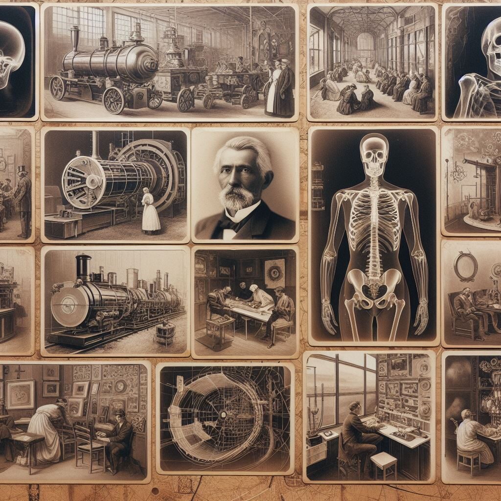
In the ever-evolving landscape of medical technology, few inventions have revolutionized diagnostic imaging as profoundly as X-ray tubes. These remarkable devices serve as the backbone of X-ray imaging, allowing healthcare professionals to peer inside the human body with unprecedented clarity and precision. Join us on an immersive journey as we delve into the intricate world of X-ray tubes, uncovering their origins, mechanics, applications, and impact on modern healthcare.

1. Origins and Evolution
The Genesis of X-Ray Technology
Our journey begins with a look back at the origins of X-ray technology. Discovered by Wilhelm Roentgen in 1895, X-rays were initially a scientific curiosity, but their potential for medical imaging was soon realized. Roentgen’s groundbreaking discovery paved the way for the development of X-ray tubes, the cornerstone of X-ray imaging.
The Evolution of X-Ray Tubes
Over the decades, X-ray tubes have undergone significant advancements and refinements. From the early Crookes tubes to modern rotating anode tubes, each iteration represents a leap forward in imaging capabilities. We explore the key milestones in the evolution of X-ray tube technology and their impact on medical diagnosis and treatment.
2. Anatomy and Mechanics
Components of an X-Ray Tube
At the heart of every X-ray tube lies a complex array of components, each playing a crucial role in the generation and emission of X-rays. We dissect the anatomy of an X-ray tube, from the cathode and anode to the glass envelope and cooling system, shedding light on the inner workings of these remarkable devices.
The Physics of X-Ray Generation
Delving deeper, we uncover the physics behind X-ray generation within the tube. Electrons emitted from the cathode are accelerated towards the anode, where they collide with target material, producing X-rays through bremsstrahlung radiation and characteristic emission. We unravel the intricacies of this process and its implications for image quality and patient safety.

3. Applications in Diagnostic Imaging
X-Ray Imaging Modalities
X-ray tubes form the backbone of a wide range of diagnostic imaging modalities, including radiography, fluoroscopy, and computed tomography (CT). We explore each modality in detail, highlighting the unique capabilities and applications of X-ray imaging in clinical practice.
Emerging Trends and Innovations
In recent years, advancements in X-ray tube technology have opened up new frontiers in diagnostic imaging. From digital radiography to cone-beam CT, we examine the latest trends and innovations shaping the future of X-ray imaging and their potential impact on healthcare delivery.
4. Challenges and Considerations
Radiation Safety and Dose Management
While X-ray imaging has revolutionized medical diagnosis, it also poses risks associated with ionizing radiation exposure. We discuss strategies for radiation safety and dose management, including dose monitoring, optimization techniques, and adherence to ALARA (As Low As Reasonably Achievable) principles.
Maintenance and Quality Assurance
Ensuring the optimal performance of X-ray tubes requires regular maintenance and quality assurance measures. We explore the importance of routine inspections, calibration, and troubleshooting to prevent downtime and maintain image quality in clinical practice.
Conclusion
Looking ahead, we glimpse into the future of X-ray tube technology and its potential impact on healthcare. From advances in material science to the integration of artificial intelligence, we speculate on the possibilities for further innovation and improvement in diagnostic imaging.
As we conclude our journey into the world of X-ray tubes, we reflect on their profound impact on modern medicine. From their humble beginnings to their pivotal role in diagnostic imaging, X-ray tubes have transformed healthcare delivery, enabling clinicians to diagnose and treat a wide range of conditions with unprecedented precision and efficiency. As technology continues to evolve, we remain optimistic about the future of X-ray imaging and its potential to improve patient care and outcomes.

